Deep Vein Thrombosis (DVT) - Ultrasound Scanning Technique
What Happens During an Arterial Leg Ultrasound?
Ultrasound Tutorial: DVT / Lower Limb Veins | Radiology Nation
Acute and chronic Deep vein thrombosis. Vein diameter variation | Ultrasound
How To Lower Extremity Ultrasound
Saphenous Vein Doppler Ultrasound Normal Vs Abnormal | Varicose Veins | Lower Limb Vascular USG
Vscan Protocol Venous Doppler Lower Extremity
Understanding Doppler Waveforms and Sounds Using the DMX
Venous ultrasound course: Deciphering DVT waveforms
Ultrasound Scan of Your Leg - The Vein Institute
Femoral Vein Doppler Ultrasound Probe Positioning | Lower Limb Veins USG Scanning Technique
Part 1: Varicose veins ultrasound assessment: GSV anatomy assessment
Varicose Veins Doppler Ultrasound Report Example | Lower Limb Venous Insufficiency Sonography USG
Dr Asif Momin - Workshop of Color Doppler of Venous System || Sonobuzz 2022
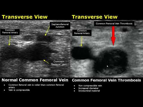
Femoral Vein Doppler Ultrasound Normal Vs Abnormal Image Appearances | Deep Vein Thrombosis USG Scan
Popliteal Deep Venous Thrombosis - Ultrasound Image Interpretation
Lower Extremity Venous Duplex Ultrasound Chronic Venous Insufficiency
Popliteal Vein Doppler Ultrasound Normal Vs Abnormal Image Appearances | Deep Vein Thrombosis USG
Portal Vein Color & Spectral Doppler Ultrasound Normal Vs Abnormal Images | Liver Vascular USG Scan
Femoral Artery Doppler Ultrasound Normal Vs Abnormal | Stenosis/Occlusion/Pseudoaneurysm/AVF USG