What Happens During an Arterial Leg Ultrasound?
Deep Vein Thrombosis (DVT) - Ultrasound Scanning Technique
Ultrasound Scan of Your Leg - The Vein Institute
Understanding Doppler Waveforms and Sounds Using the DMX
What is the significance of Doppler study for legs? - Dr. Surekha Tiwari
Ultrasound Tutorial: DVT / Lower Limb Veins | Radiology Nation
Femoral Artery Doppler Ultrasound Normal Vs Abnormal | Stenosis/Occlusion/Pseudoaneurysm/AVF USG
Popliteal Vein Doppler Ultrasound Normal Vs Abnormal Image Appearances | Deep Vein Thrombosis USG
Acute and chronic Deep vein thrombosis. Vein diameter variation | Ultrasound
How To Lower Extremity Ultrasound
Color Doppler of Leg | पैरों का Ultrasound कैसे होता है?
Using a Doppler
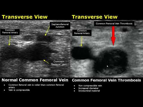
Femoral Vein Doppler Ultrasound Normal Vs Abnormal Image Appearances | Deep Vein Thrombosis USG Scan
How To Lower Extremity Arterial Duplex Exam
Femoral Vein Doppler Ultrasound Probe Positioning | Lower Limb Veins USG Scanning Technique
PAD vs PVI cartoon animation & memory tricks peripheral arterial disease pathophysiology, signs
Doppler Ultrasound Test | Doppler Ultrasonography |
Part 1: Varicose veins ultrasound assessment: GSV anatomy assessment
Popliteal Deep Venous Thrombosis - Ultrasound Image Interpretation
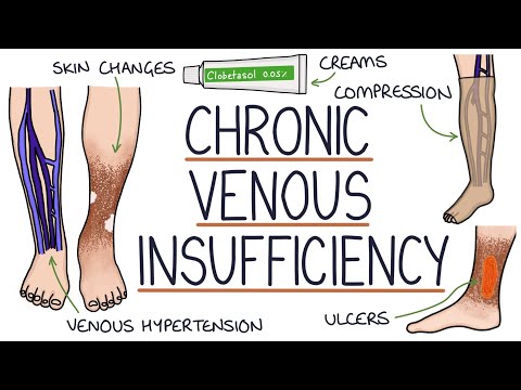
Understanding Chronic Venous Insufficiency